Diagnostic Imaging: X-ray, bone scan, MRI, ultrasound. Which test is the right choice to diagnose your problem?
Posted June 27th, 2008

The first Nobel Prize in physics went, in 1901, to a German scientist named Wilhelm Roentgen for his work in the development of the x-ray. He completed his work in 1895, but it was just the beginning of the road for diagnosing illness and injury with the creation of images inside the human body. Diagnosis established with imaging techniques is one of the most important tools in medicine today.
X-ray
Modern x-ray, also called radiography, has little in common with Roentgen’s images or even films that were exposed a decade ago. Today, x-ray can be all digital. This means the clarity is unbelievably sharp, and the clinician can manipulate and enhance the pictures for more precise interpretation. The entire process is completed surprisingly fast. When needed, radiology consultations can be gathered quickly via electronic communications systems.
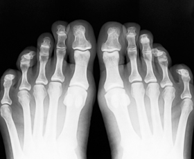
X-rays are a form of electromagnetic radiation, just like visible light. These particles pass through the body, and a computer or special film is used to record the images. Dense structures such as bone will block most of the x-ray particles, and will appear white. Structures containing air will be black, and muscle, fat, and fluid will appear as shades of gray.
X-rays should be first in line for diagnosing athletic injuries. It is inexpensive, safe, and readily available. Coupled with good clinical skills, the x-ray will often solidify a diagnosis. The foot and ankle are complicated anatomical structures, and an x-ray is often used to assess anatomical abnormalities in the foot and leg that may be an issue for the repetitive motion athlete.
Bone Scans
A bone scan is a test that detects areas of increased or decreased bone metabolism. It is performed to identify abnormal processes involving the bone, such as tumor, infection, or fracture.
A radiotracer (a bone-seeking radioactive material) is injected into a vein, so it travels through the bloodstream. As the material wears away, it gives off radiation. This radiation is detected by a camera that slowly scans the body. The camera takes pictures of how much radiotracer collects in the bones.
X-rays will not show a stress fracture, but a bone scan will. This exam is not as expensive as an MRI and offers more conveniences with time and availability. The limitation of this exam, especially with the running athlete, is false positive diagnosis. The bone scan does not produce actual pictures that can be interpreted and viewed like an x-ray. Rather, it will demonstrate blood flow that can indicate an injury or if a healing process has occurred. A runner often has “hot spots” in legs and feet because of the repeated motion and increased gravitational forces and stress on the lower extremity. These spots can be falsely interpreted as an area of stress fracture. For this reason, careful clinical correlation is important when the bone scan is used as a diagnostic tool.
MRI
Many providers in sports medicine are turning to Magnetic Resonance Imaging for diagnosis of stress-related bone injury, as this modality, unlike the bone scan, is very precise. Over the last few years, radiologists have been able to determine stress reactive tissues in bone that can foretell of a stress fracture injury about to happen.
The MRI is in a class alone when it comes to soft tissue injury. Tendons, joint structures, nerves, and blood vessels can all be seen easily and distinctly. Consider the MRI as a complete full thickness visual, from one side to the other, of any anatomical part. The MRI is not a continuous film-like process, but very small slices of imagery seamed together. This does create a small flaw in the magic of the exam. An example is the interdigital neuroma in the foot. The neuroma and nerve compressions may be so small that they can fall between slices of exposure, creating false readings. Since the MRI relies on a strong magnetic field, it cannot be used when many of the metals used for fracture or joint repairs are surgically implanted.
The MRI is an expensive exam. Be sure that your medical insurance has authorized the event, or you may be saddled with a rather large bill.
Ultrasound
An ultrasound machine creates images that examine body organs by sending out high-frequency sound waves, which reflect off body structures. A computer receives these reflected waves and uses them to create a picture. Unlike with an x-ray, there is no ionizing radiation exposure with this test.
Most commonly known for fetal exams during the course of pregnancy, the ultrasound has many other applications that can be helpful in diagnosis and treatment of athletic injuries. Ultrasound is generally inexpensive compared to other modalities. At one time, ultrasound machines were only found in hospitals, but today, many large facilities that treat athletes will have the equipment and necessary training to use it. Ultrasound is being used with increased frequency for trigger point injection therapy. The images make it possible to precisely select small anatomical structures, guiding the provider to inject specifically and accurately medications that can treat injury and relieve pain. Office-based use of ultrasound increases successful treatment programs in athletes.
Summary
The answer to accurate diagnosis is often locked inside a picture. Roentgen must have known this when he found the key to imaging the inside of the human body. Radiologists and biomechanical scientists have an ongoing dedication to interpreting images. The x-ray should be the first and most basic exam performed for almost all athletic foot and leg injuries. Interpretation should always be coupled with accurate, detailed clinical findings.
